If your child suffers a bad blow to the head or experiences severe
unexplained stomach pain, landing you in the emergency department, it can
be scary. The sooner you can get answers, the sooner your fears can be
allayed, your child can get treatment and start feeling better.
As a pediatric radiologist, I use imaging scans to get those answers
quickly, which can make all the difference in the middle of a
life-threatening emergency. But I recognize that the benefits of these
tests, some of which use radiation, must be balanced with potential risks
for children. Exposure to ionizing radiation is a risk factor for
developing cancer later in life. The effects of radiation are cumulative,
so the radiation a child receives today will be with him or her forever.
Moreover, it’s important to remember that with diagnostic testing, as well
as with treatment, kids are growing and their tissue is more sensitive.
The most common imaging scan that uses radiation is digital radiography,
more commonly known as an X-ray. Radiography uses X-rays, radiant energy
beams like light or radio waves that pass through the body, to produce
images of the body’s internal structures, such as bones, organs and teeth.
A single X-ray, such as one used to image a child’s fractured ankle, uses
very little radiation, and the benefits of the test typically far outweigh
the risks. We also use lead aprons to cover the parts of the body not being
imaged to further limit the child’s exposure.
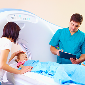
For more detailed imaging, computed tomography (CT) has proven to be a
useful tool. First developed in the 1970s, CT combines hundreds of X-rays
taken from many angles to produce detailed cross-sectional images of one
part of the body. Largely because these scans are fast, accurate and
non-invasive, they are used 10 times more now than they were in 1980. In
the United States alone, between 5 and 9 million CT scans are performed
annually on children.
CT scans are often used to evaluate a head injury or abdominal pain in
children, but CT scans can also help us diagnose fractures and internal
injuries, as well as cancers, infections and blood clots. There are clear
benefits to CT scans: When we find a cancerous tumor early before it’s had
time to spread, for example, the scan can be lifesaving.
However, because CT scans expose patients to several hundred times more
radiation than an X-ray, those benefits must be weighed very carefully
against the risk of exposure. Nearly half of children in emergency
departments with a head injury end up getting a CT scan, according to the
American Academy of Pediatrics, and one-third of these scans are not
necessary. For children with mild concussions, CT scans usually come back
normal exposing the child to radiation while providing no clinical benefit.
These scans are much more useful for bleeding in the brain and skull
fractures.
This is why it’s important for a physician to ask detailed questions about
the injury and symptoms before considering what tests to run. Whenever
possible, I always first try to find answers with an MRI or an ultrasound –
neither of which produce radiation (MRIs use radio waves and ultrasound
uses sound waves to create an image). Yet, CT scans are sometimes the best
modality of imaging in certain conditions, so much depends on the child and
his or her condition.
There are ways to minimize a child’s exposure to radiation without forgoing
the benefit of this powerful tool when it’s truly needed.
First, I recommend that before consenting to a CT scan for a child, parents
should ask their doctor about using alternative forms of imaging that do
not involve radiation.
Second, it’s wise to confirm that the facility’s CT scan settings can be
adjusted for the child’s size, so that your child does not receive a higher
radiation dose than is truly necessary. Fortunately, it’s common for
emergency departments, as well as most other imaging centers, to be
equipped with pediatric dose protocols for CT scans.
Many facilities, like the ones we use at Kaiser Permanente, have specific
protocols for children, including equipment capable of low-dose CT
scanning. Our scanners have built-in protocols with a type of software that
allows us to make adjustments to the scanner according to the size and age
of the child. Depending on the test being performed, these scanners can
reduce the dose for a baby or child by as much as half of what an adult
would receive.
Third, ask if the CT technologist can scan only the part of the body that
needs to be examined. If your child has abdominal pain and doctors suspect
appendicitis, for example, the radiologist can scan only the lower abdomen
and pelvis, excluding the lower chest and upper abdomen. Limiting the scan
to the affected area lowers the overall radiation dose.
Finally, confirm that the radiologist has saved the dose information for
each scan, so that the amount of radiation your child has been exposed to
is documented and can be used when considering future scans. Information on
radiation dose is often automatically saved by the equipment into the
imaging database.
Taking these simple steps can reduce the risk of radiation exposure in your
child – which also reduces the possible slight risk of your child
developing a future radiation-related cancer. But parents should also keep
in mind that, while no amount of radiation is considered absolutely safe,
the cancer risks associated with imaging scans are small. We just need to
minimize the risks for kids whenever possible.
For more guidance on safe and effective medical imaging for children,
visit the Parent section of
imagegently.org. For information about dental imaging, visit the American Academy of Pediatric Dentistry site.


